
Vitrectomy
 The vitreous is the clear, jelly-like gel that fills the middle of the eye. Opacities in the vitreous can cause symptomatic floaters or reduced vision. The vitreous gel is attached to the retina in our youth but it is normal for the gel to pull away from the retina later in life. This pulling away from the retina has the potential to negatively impact the retina and is the most common cause of retinal tears and detachments.
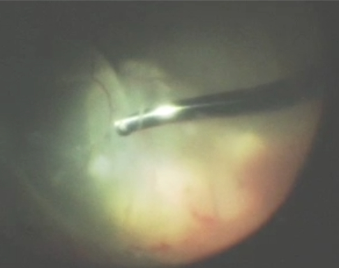
A vitrectomy is an outpatient surgical procedure that is the best way to treat certain ocular conditions. An instrument called a vitrector (see image) is used to cut and suction out the vitreous. Once the vitreous is removed, it never grows back. Instead, fluid in the front part of the eye called aqueous fluid will fill up the space where the vitreous was removed. The eye does not need vitreous to be healthy – sometimes the vitreous is referred to as the appendix of the eye in that it causes nothing but problems for the patient!
Ocular conditions that may benefit from treatment with vitrectomy include: retinal detachment, diabetic retinopathy, endophthalmitis (eye infection), macular hole, macular pucker/epiretinal membrane, vitreous hemorrhage.
A vitrectomy is considered a safe procedure, but there are risks to any surgery. Some of the risks include intraocular bleeding, infection, swelling of the retina, retinal tears or detachments, cataract formation, or scar tissue formation.