Fluorescein Angiography (FA)
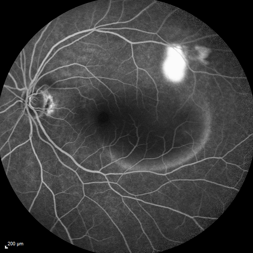
 In order to find abnormal blood vessels under the retina and/or to identify conditions that can cause retinal swelling and reduced vision, it is sometimes necessary to perform a test called angiography.
This test is performed by injecting a dye into the vein of the arm, then photographing the dye as it passes through the circulation in the back of the eye. Depending on the pattern of dye transmission and leakage, certain disease processes can be identified. Two different dyes are commonly used: fluorescein and indocyanine green. Special digital cameras joined to computers are used to maximize the effectiveness of this test.
Doctors choose fluorescein angiography to study diseases of the retinal and choroidal blood vessels within the eye. The results of this test enable the physician to diagnosis many abnormalities of the retina and choroid that could not be diagnosed accurately otherwise. The results of this study also serve as a guide to laser treatment for many diseases of the retina and choroid.


