Lattice Degeneration
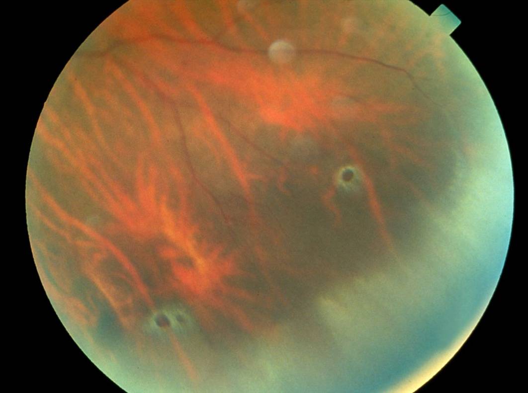
Lattice degeneration is a thinning of the peripheral retina that sometimes is associated with retinal holes.
Lattice degeneration occurs in about 8% of the general population and is more common in eyes that are nearsighted (myopic).
The patient usually does not have symptoms from lattice, and often no treatment is required. However, because lattice is a thinning of the retina and retinal tears and detachments are more likely to originate in these areas, prophylactic laser treatment may be considered — especially if the patient experiences flashing lights, new floaters, or a curtain-like shade over their vision, which are retinal detachment warning signs. In addition, laser may be recommended to an eye with lattice in a patient with a history of retinal detachment in the other eye.
Laser treatment is performed in the office. A numbing drop is applied to the eye and a contact lens may be used to focus the laser light. Then the laser is applied around the area of lattice. During laser treatment, the patient experiences bright flashes of light. After the laser treatment, vision can be blurry for up to 30 minutes, but recovers. The eye may be sore for 24 hours and the discomfort is usually relieved with Tylenol or Advil. The laser spots induce small scars to form that adhere the retina to the eye wall, acting as a seal or “glue” in the area of the lattice that will reduce the risk of future retinal detachment.


